Simulation-Based Algorithm Design: Deep Learning or Machine Learning for Finding the Number of Stones in CT Scan/MRI of Kidney
DOI:
https://doi.org/10.62497/irjai.145Keywords:
Kidney stones, Nephrolithiasis, CT imaging, MRI imaging, Deep learning, Machine learning, Clinical decision support, Hybrid algorithmAbstract
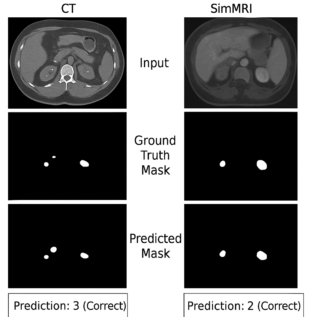
Background: Kidney stone disease (nephrolithiasis) is a prevalent urological condition. While modern imaging modalities such as CT and MRI enable rapid detection and localization of stones, automatically estimating stone counts remains challenging due to variations in size, contrast, resolution, and anatomical positioning. Objective: To develop and evaluate a multimodal algorithm that enhances the accuracy and robustness of automatic kidney stone detection and counting across CT and MRI imaging. Methods: A hybrid framework, StoneNet-HC, was designed, combining a lightweight convolutional neural network (TinyResNet-FeatureNet) for stone region detection with a Random Forest regression model for predicting stone counts. The approach incorporated multimodal datasets, including publicly available CT scans and synthetically generated MRI images simulating low-contrast conditions. Synthetic augmentation techniques were applied to improve generalizability. Performance was assessed against existing methods using mean absolute error (MAE), Dice coefficient, Intersection over Union (IoU), and classification accuracy. Results: StoneNet-HC achieved lower MAE, improved Dice and IoU scores, and higher classification accuracy compared to state-of-the-art approaches. The system demonstrated consistent performance across both CT and MRI modalities, showing resilience to contrast variability and resolution differences. Conclusion: This study presents a simulation-driven, hybrid algorithm that integrates deep learning detection with machine learning regression to enable accurate and generalizable kidney stone quantification. The modular design supports potential integration into clinical diagnostic workflows, bridging high accuracy with improved interpretability for multimodal imaging analysis.
Downloads
References
. Scales CD, Smith AC, Hanley JM, Saigal CS, Urologic Diseases in America Project. Prevalence of kidney stones in the United States. European urology. 2012 Jul 1;62(1):160-5. https://www.sciencedirect.com/science/article/abs/pii/S0302283812004046
. Fulgham PF. Clinical Effectiveness Protocols for Imaging in the Management of Ureteral Calculus Disease. AUANews. 2012 Apr 1;17(4). https://doi.org/10.1016/j.juro.2012.10.031
. Türk C, Petřík A, Sarica K, Seitz C, Skolarikos A, Straub M, Knoll T. EAU guidelines on interventional treatment for urolithiasis. European urology. 2016 Mar 1;69(3):475-82. https://www.sciencedirect.com/science/article/abs/pii/S0302283815007009
. Litjens G, Kooi T, Bejnordi BE, Setio AA, Ciompi F, Ghafoorian M, Van Der Laak JA, Van Ginneken B, Sánchez CI. A survey on deep learning in medical image analysis. Medical image analysis. 2017 Dec 1;42:60-88. https://www.sciencedirect.com/science/article/abs/pii/S1361841517301135
. Lundervold AS, Lundervold A. An overview of deep learning in medical imaging focusing on MRI. Zeitschrift fuer medizinische Physik. 2019 May 1;29(2):102-27. https://www.sciencedirect.com/science/article/pii/S0939388918301181
. Alzubaidi L, Zhang J, Humaidi AJ, et al. Review of deep learning: concepts, CNN architectures, challenges, applications, future directions. J Big Data. 2021; 8(53). https://doi.org/10.1186/s40537-021-00444-8
. Hoeser T, Kuenzer C. Object detection and image segmentation with deep learning on earth observation data: A review-part i: Evolution and recent trends. Remote Sensing. 2020 May 22;12(10):1667. https://doi.org/10.3390/rs12101667
. Cui Y, Sun Z, Ma S, Liu W, Wang X, Zhang X, Wang X. Automatic detection and scoring of kidney stones on noncontrast CT images using STONE nephrolithometry: combined deep learning and thresholding methods. Molecular Imaging and Biology. 2021 Jun;23(3):436-45. https://link.springer.com/article/10.1007/s11307-020-01554-0
. Sharma, S., Choudhury, B., & Kaushik, A.. Classification of urinary stone images using machine learning algorithms: A comparative study. International Journal of Computer Applications, 184(8), 9–14. https://doi.org/10.5120/ijca2022912256
. Yildirim, E. A., Aydin, Z. Y., & Tasci, B. Deep learning-based kidney stone detection on CT images. Computerized Medical Imaging and Graphics, 91, 101944. https://doi.org/10.1016/j.compmedimag.2021.101944
. Sheafor DH, Hertzberg BS, Freed KS, Carroll BA, Keogan MT, Paulson EK, DeLong DM, Nelson RC. Nonenhanced helical CT and US in the emergency evaluation of patients with renal colic: prospective comparison. Radiology. 2000 Dec;217(3):792-7. https://pubs.rsna.org/doi/abs/10.1148/radiology.217.3.r00dc41792
. Shokeir AA, Abdulmaaboud M, Farage Y, El-Nahas AR. Comparison of unenhanced helical CT and magnetic resonance urography in urinary tract obstruction. Journal of Urology; 2010 183(3),952–955. https://doi.org/10.1016/j.juro.2009.11.017
. Çiçek Ö, Abdulkadir A, Lienkamp SS, Brox T, Ronneberger O. 3D U-Net: learning dense volumetric segmentation from sparse annotation. InInternational conference on medical image computing and computer-assisted intervention 2016 Oct 2 (pp. 424-432). Cham: Springer International Publishing. https://link.springer.com/chapter/10.1007/978-3-319-46723-8_49
. Chen H, Dou Q, Yu L, Qin J, Heng PA. VoxResNet: Deep voxelwise residual networks for brain segmentation from 3D MR images. NeuroImage. 2018 Apr 15;170:446-55. https://www.sciencedirect.com/science/article/abs/pii/S1053811917303348
. Krizhevsky A, Sutskever I, Hinton GE. Imagenet classification with deep convolutional neural networks. Advances in neural information processing systems. 2012;25. https://proceedings.neurips.cc/paper/2012/hash/c399862d3b9d6b76c8436e924a68c45b-Abstract.html
. Simonyan K, Zisserman A. Very deep convolutional networks for large-scale image recognition. arXiv preprint arXiv:1409.1556. 2014 Sep 4. https://arxiv.org/abs/1409.1556
. He K, Zhang X, Ren S, Sun J. Deep residual learning for image recognition. InProceedings of the IEEE conference on computer vision and pattern recognition 2016 (pp. 770-778). https://www.cv-foundation.org/openaccess/content_cvpr_2016/papers/He_Deep_Residual_Learning_CVPR_2016_paper.pdf
. Redmon J, Farhadi AY. An incremental improvement. arXiv preprint arXiv:1804.02767. 2018 Apr 8. https://ask.qcloudimg.com/draft/2661027/i16j8zgndj.pdf
. Hatamizadeh A, Tang Y, Nath V, Yang D, Myronenko A, Landman B, Roth HR, Xu D. Unetr: Transformers for 3d medical image segmentation. InProceedings of the IEEE/CVF winter conference on applications of computer vision 2022 (pp. 574-584). https://openaccess.thecvf.com/content/WACV2022/html/Hatamizadeh_UNETR_Transformers_for_3D_Medical_Image_Segmentation_WACV_2022_paper.html
. Selvaraju RR, Cogswell M, Das A, Vedantam R, Parikh D, Batra D. Grad-cam: Visual explanations from deep networks via gradient-based localization. InProceedings of the IEEE international conference on computer vision 2017 (pp. 618-626). https://openaccess.thecvf.com/content_iccv_2017/html/Selvaraju_Grad-CAM_Visual_Explanations_ICCV_2017_paper.html
. Wang M, Deng W. Deep visual domain adaptation: A survey. Neurocomputing. 2018 Oct 27;312:135-53. https://www.sciencedirect.com/science/article/abs/pii/S0925231218306684
. Lundberg SM, Lee SI. A unified approach to interpreting model predictions. Advances in neural information processing systems. 2017;30. https://proceedings.neurips.cc/paper/2017/hash/8a20a8621978632d76c43dfd28b67767-Abstract.html
. Dosovitskiy A, Beyer L, Kolesnikov A, Weissenborn D, Zhai X, Unterthiner T, Dehghani M, Minderer M, Heigold G, Gelly S, Uszkoreit J. An image is worth 16x16 words: Transformers for image recognition at scale. arXiv preprint arXiv:2010.11929. 2020 Oct 22. https://arxiv.org/pdf/2010.11929/1000
. Perez L, Wang J. The effectiveness of data augmentation in image classification using deep learning. arXiv preprint arXiv:1712.04621. 2017 Dec 13. https://arxiv.org/abs/1712.04621

Downloads
Published
Issue
Section
License
Copyright (c) 2024 Zia Ur Rehman (Author)

This work is licensed under a Creative Commons Attribution-NonCommercial 4.0 International License.
